| |

![]() Stroke is the third leading cause of
death in this country.
The major cause of stroke is disease in the carotid arteries, which supply blood to the brain. Stroke from hardening of the arteries (atherosclerosis) may be prevented by surgical
intervention (carotid endarterectomy). At Stony Brook, our goal is to reduce the number of strokes in our patients.
Stroke is the third leading cause of
death in this country.
The major cause of stroke is disease in the carotid arteries, which supply blood to the brain. Stroke from hardening of the arteries (atherosclerosis) may be prevented by surgical
intervention (carotid endarterectomy). At Stony Brook, our goal is to reduce the number of strokes in our patients.
The term stroke actually refers to a large range of diseases, the usual result of which is the sudden onset of symptoms; from mild weakness of an arm or leg to loss of speech, paralysis, coma, and death. Many people with this disease have no symptoms. A serious problem, TIAs (transient ischemic attacks, often called "mini-strokes") are strokes, but the symptoms last less than 24 hours.
A TIA is a big warning that a major stroke is in your future. This is not something to take lightly. Although these events are often called "mini-strokes" and shrugged off by patients and even some doctors, they should not be ignored. For 1 in 3 people, a TIA will result in a full-blown stroke within five years, if left untreated. Usually the strokes occur within a few days or a weeks of the TIA. The buildup of plaques in the carotid or vertebral arteries happens over time, and there are usually no symptoms until the narrowing reaches a critical state.
 For an excellent online resource on stroke, see the American Heart Association's Heart and Stroke Encyclopedia.
Peripheral vascular disease refers to diseases of any of the
blood vessels outside of the heart and to diseases of the
lymph vessels. It is often a narrowing of the blood vessels
that carry blood to leg and arm muscles. There are two
types of these circulation disorders: Functional Peripheral Vascular Disease These diseases are not organic in cause and do not
involve defects in the structure of the blood vessels.
They are short-term effects and may be reversed.
An example is Raynaud’s disease or phenomenon, a
condition in which the smallest arteries that bring blood
to the fingers or toes constrict (go into spasm) when
exposed to cold or as the result of emotional upset.
This disease happens most commonly in women
between the ages of 18 and 30. Organic Peripheral Vascular Disease These diseases are caused by structural changes, such
inflammation and tissue damage, in the blood vessels.
An example is Buerger’s disease (thromboangiitis
obliterans), a chronic inflammatory disease found
chiefly in the peripheral arteries and veins of the
extremities. This disease most commonly happens to
men between the ages of 20 and 40 who smoke
cigarettes. Symptoms include pain in the legs or feet,
clammy and cold skin, and a diminished sense of heat
and cold. Aside from the size, there are other things our physicians look for as an indication that surgery is
needed. If a patient presents with the "blue toe" syndrome, a sign that emboli
(bits of plaque) have been sent to the foot or leg, surgical
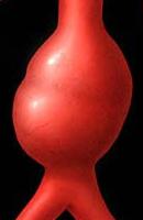
intervention is apt to be discussed. Dissecting aneurysm occurs when blood gets through a lengthwise tear between layers of the wall
of an artery. These layers
then separate and swell, making a balloon-shaped formation that causes severe pain.
This condition—which can be fatal if the artery bursts—can be caused by a disease, birth defect, or injury. It is usually caused by
hardening of the arteries (atherosclerosis), a common disorder that causes narrowing of arteries and reduces
circulation. High blood pressure also contributes to this disease.
Patients with thoracic dissecting aneurysms describe the pain in the chest as being of a "ripping" or "tearing"
character, which is frequently mistaken for a heart attack. The preferred treatment for dissecting aneurysm is immediate surgery. The surgeon replaces the weakened
part of the artery with a graft made of artificial material. If patients have high blood pressure,
they are generally given fast-acting intravenous medication to lower it. Unless it is surgically removed, an aneurysm is a permanent condition. The outlook
varies, depending on the location of the aneurysm and the individual patient's age and overall health. The
less urgent the need for surgery, the better the chance for survival. Varicose veins are blood vessels, usually in the legs, that become
permanently dilated (widened) and twisted. They may include
superficial veins, deep veins and veins that connect superficial
and deep veins. The signs of varicose veins are
enlarged, disfiguring, snakelike, bluish veins which are
visible under the skin upon standing; they appear most
often in the back of the calf or on the inside of the leg
from ankle to groin. Symptoms may include vague discomfort and aching in the legs, especially
after standing; and fatigue. Click here to see photos of varicose veins in the leg before and after treatment. Spider veins—for which the medical term is telangiectasias—are
tiny, superficial blood vessels which have become
permanently dilated. The veins are generally blue,
red, or purple in color. Although spider veins can occur on
most areas of the body, common sites for the condition are
the legs and face.
Up to 41% of women and 15% of men in the
United States have spider veins. The veins usually cause no
serious problems, but some patients experience burning or
pain in the affected area of the body. For many people,
especially those with large areas of spider veins, the condition
can be cosmetically unappealing. Click here to see photos of spider veins in the leg before and after treatment.